Showing posts with label asthma. Show all posts

15 May, 2009

Breast Milk Transfer of Antigens Establishes Anti-Allergenic Tolerance to Those Antigens

ResearchBlogging.orgMilk isn't just milk. The pasteurized cow milk that we can purchase in the grocery store has been cleaned, processed, and in many cases chemically scrubbed of fat. Unique among mammals, we Western humans stubbornly persist in our consumption of dairy products well into adulthood regardless of whether or not our guts like it. But the cow milk in the store is very different from the fresh milk humans nurse their newborns with. Fresh human milk contains growth factors, vitamins, an astounding density of calories, IgA antibodies, and also antigen. Disregarding the controversial and impassioned debate surrounding breast milk vs. infant formula, research has found that the immunological molecules secreted in breast milk are important for the developing immune system of the infant.

Verhasselt and Julia et al have demonstrated that antigen secreted in a mother's breast milk significantly impacts the later development of allergic asthma in her infants. In effect, this is immunological programming.

They used an elegant experimental system wherein they took nursing dams (mouse mother) and exposed them to antigens (OVA) without their pups, then placed them back with their pups to nurse. Later on when the pups had reached adulthood, they sensitized the mice per the immunology canon and tested their allergic asthmatic (hereafter refered to as atopic) response. In mice breastfed by OVA-exposed dams it was found that, in comparison to those breastfed by non-exposed dams, airway hyperreactivity, pulmonary eosinophilia, cellular infiltrate, and mucus deposition were all decreased towards mice not challenged with OVA (normal, negative controls). Moreover, the classical TH2 cytokines (IL-4, IL-5, IL-10, and IL-13) that have been associated widely with atopic responses were decreased in OVA-breastfed mice, as were the frequencies of the lung CD4+ T-cells that secrete them. Overall, this points to OVA-breastfed mice having a significantly weakened allergenic response to an allergen, or put another way, these mice tolerated the prescence of the allergen much better.

As an aside, OVA is an abbreviation for ovalbumin, which is a key molecule in eggs and is a very sticky molecule. In every study of allergy or asthma that I can remember reading, OVA was used to sensitize mice and produce an allergic response to it. This is done by injecting a solution of OVA into the peritoneal cavity of the mice, waiting 2 weeks to let OVA-specific CD4+ T-cells develop, and then challenging the mice by squirting an OVA aerosol up their noses. This model has proven to reliably produce a strong and specific allergenic response.

But how did the authors determine that it was secreted antigen itself that was inducing the tolerance?

To address this, they took normal, wild-type C57 mouse pups and gave them to lactating uMT and RAG-2 transgenic dams*. Both these transgenic strains of mice are completely unable to mount an adaptive immune response to anything; they do not and cannot make antibodies. When these mice were exposed to OVA and then nursed the wild-type pups, the same effects of the OVA-breastfeeding inducing allergen tolerance were observed. This was also replicated with Balb/c mice (which is important because the C57 strain is known to skew towards a TH1 response phenotype while Balb/c skews more to TH2, which is better characterized in the pathophysiology of atopy).

But then Verhasselt and Julia took a look at some important and specific immunoregulatory molecules: TGFb and IL-10. Both of these molecules are broad suppresors of inflammatory immune responses regardless of that response's cellular phenotype. IL-10 transgenic dams did not alter observed results when exposed to OVA, but TGFb knockdown dams did in that the pups they nursed were reactive to OVA. This strongly suggests that TGFb must accompany antigen in the breast milk in order for the infants' immune systems to recognize that antigen as a harmless allergen and, in effect, program itself not to react against it. Luckily for human mothers, though, breast milk already contains TGFb.

At a wider level, this suggests that mothers who are exposed to many allergens will pass on tolerance to those allergens to any breast-feeding infants they may have. Perhaps this, coupled with the hygiene hypothesis, is a call for more moms to teach their infants how to make mud pies even earlier.

*I can't help but wonder how many times they did this and found all the pups had been killed off by their adoptive mothers, as this is what stressed out rodent dams tend to do.

Verhasselt, V., Milcent, V., Cazareth, J., Kanda, A., Fleury, S., Dombrowicz, D., Glaichenhaus, N., & Julia, V. (2008). Breast milk–mediated transfer of an antigen induces tolerance and protection from allergic asthma Nature Medicine, 14 (2), 170-175 DOI: 10.1038/nm1718

25 March, 2009

Not-So-Mad Science: IL-13 vs. IL-4 In The Battle For Asthma!

ResearchBlogging.org(Previous asthma research-blogging here)

Marsha Wills-Karp, Jackie Luyimbazi, Xueying Xu, Brian Schofield, Tamlyn Y. Neben, Christopher L. Karp, Debra D. Donaldson (1998). Interleukin-13: Central Mediator of Allergic Asthma Science, 282, 2258-2261

Abstract:
The worldwide incidence, morbidity, and mortality of allergic asthma are increasing. The pathophysiological features of allergic asthma are thought to result from the aberrant expansion of CD4(+) T cells producing the type 2 cytokines interleukin-4 (IL-4) and IL-5, although a necessary role for these cytokines in allergic asthma has not been demonstrable. The type 2 cytokine IL-13, which shares a receptor component and signaling pathways with IL-4, was found to be necessary and sufficient for the expression of allergic asthma. IL-13 induces the pathophysiological features of asthma in a manner that is independent of immunoglobulin E and eosinophils. Thus, IL-13 is critical to allergen-induced asthma but operates through mechanisms other than those that are classically implicated in allergic responses.

There are many morbidly fascinating pathological changes associated with onset and clinical asthma. To wit, these include eosinophilia, mucus overproduction, mast cell/other inflammatory cell airway infiltration, and increased smooth muscle. There may also be scarring of the airways.

From what I currently understand about it, airway hypersensitivity generally happens after immune effector cells have infiltrated the underlying airway tissues. When these effector cells, which can include allergen-specific T-cells, mast cells, eosinophils, basophils, and even macrophages, are activated by an irritant (the allergen) they more or less cut loose and let wild with the localized inflammation. The localized inflammation, in turn, leads to more immune cell infiltration over time and concurrently the tissue itself undergoes histopathologically apparent changes, including thickening of the base layer of smooth muscle.

For example of immune effector cells getting activated, let's consider the most dramatic case: the mast cell. Mast cells are a type of white blood cell that expresses Fc receptors for IgE (IgE is the immunoglobulin most associated with allergic and anti-parasite responses) on it's surface. The Fc-bound IgE act as allergen-specific receptors that, when bound to their ligand, send a signal into the cell to degranulate. Mast cells store relatively massive amounts of inflammatory cytokines and peptides in large granules (e.g., histamines, prostaglandins, and leukotrienes) and they can, effectively, disgorge them all at once. This can lead to a very rapid spike in the systemic concentration of inflammatory effector molecules and subsequently extremely rapid onset of asthmatic symptoms. The same process is at work in acute food allergies.

Figure A: The mast cell is the one with the big lumpy nucleus in the center. The black dots surrounding it are granules packed with inflammatory molecules, just waiting to be released and wreak havoc. Those other 2 cells to the right are lymphocytes (according to the original caption on this TEM).

But what inflammatory molecules are required to invoke and/or sustain a hypersensitive airway response?

This paper examined the role of IL-13 in allergic asthma. According to Janeway's Immunobiology, IL-13 is involved in the differentiation of naive CD4+ T-cells into TH2 cells, which have been shown to be more involved in allergy than TH1 cells. IL-13 is also secreted by TH2 cells, apparently, and has been shown to have a direct effect on airway epithelial cells by which their proliferation in increased and differentiation into goblet cells (goblet cell metaplasia) is increased, which in turn leads to the increased mucus production seen in allergy and asthma. And when your organs are infected with multicellular parasites, IL-13 is there to help the organs make the changes they need to get rid of those parasites. And as if that weren't enough, IL-13 also increases smooth muscle contractility.

But IL-13 doesn't really do anything without the context of a TH2 immune response. Th2 cells are characterized by secretion of IL-4, and it should be noted that IL-4 and IL-13 share a subunit in their receptors.

Figure B: The left column has a normal lung biopsy (top) and a normal airway (bottom) from a Tbet+/+ mouse. The right column has the same measurements, but showing airway inflammation with lymphocyte and eosinophil infiltration (top) and remodeled airway with increased collagen (bottom) from a Tbet-/- mouse. The picture is blurry because I took it with my phone. It is from Janeway's Immunobiology, 7th ed., page 575. Tbet is a transcription factor that is necessary for the development of TH1 cells, so its abscence will invariably result in a TH2 inflammatory response (right column). Tbet is analogous to GATA3 in TH2 cells.

Allow me to explain T-cell differentiation really briefly:
1) Naive T-cells arrive in thymus.
2) Naive T-cells have to decide whether or not to be CD4+ or CD8+, which will result in being able to recognize MHCII or MHCI, respectively.
3) CD4+ T-cells get stimulated by DCs or stuff, and the resulting cytokine mileau determines whether they become TH1, TH2, Treg (also refered to as TH3), or TH17. They can also become memory T-cells of any of those variety later on in. Respectively, these cell types are characterized by secretion of IFNg, IL-4/IL-5, IL-10, and IL-17.
4) TH1, TH2, Treg, and TH17 all more or less have distinct biological roles, although the cytokine soup that gives rise to different types is messy (e.g., IL-2 just drives T-cell proliferation irrespective of subset) and often overlaps, and they'll even compete against each other (IL-12 drives TH1 proliferation but inhibits TH2 proliferation while IL-4 does the same for TH2 cells).
So, anyway, the group behind this paper found that while IL-4 is sufficient to initiate asthmatic events, IL-13 is required for the development of the airway hypersensitivity response (AHR). They used the standard ovalbumin (OVA) induced model of AHR and found that blocking IL-13 with an neutralizing fusion protein prevents the development of AHR. Apparently blocking IL-13 in mice who already have AHR results in their measures of AHR decreasing (specifically goblet cell metaplasia and mucus production). However, with all of this, blocking IL-13 had no effect whatsoever on net circulating IgE or eosinophilia.

These findings prove that IL-13 has a significant role in asthma. But they also imply that IL-13 does not play this role through classical allergy pathways, as IL-13 is found to be elevated in patients with both allergic and non-allergic asthma. This is further supported by the group's finding that daily intratracheally administration of IL-13 is sufficient to induce asthmatic pathology even in the abscence of antigen sensitization.

What I wonder about here is: how does it make biological sense for a molecule involved heavily in the production of allergen-specific TH2 cells to also operate completely independently of that cellular phenotype?

But what is important to human health is that this paper demonstrates that adminstration of IL-13 agonists or blockers may be of great therapeutic value to human asthmatics. This paper is 11 years old, and I don't currently know if anything has come of their findings, but still, it'd be cool if this really did have therapeutic value because, as my last post on asthma discussed (link up top), inhaled acute anti-inflammatories may only be getting to the pieces of lung that need it least (because they're the pieces that can still pump air, and if reacting tissue isn't pumping tidal volume, how can inhaled medicine get to it?). If this could be used daily as a preventative, I think it could greatly improve the quality of life for asthmatics everywhere.

05 March, 2009

Not-So-Mad Science: Patchiness in Asthma

ResearchBlogging.orgIt’s a beautiful spring day, the first warm and sunny day of the year and the snow has finally melted. So naturally, you’re outside, enjoying it. The birds have reemerged from their hiding places, and all the plants are heavy with green and bloom. The whole earth smells clean, as though the snow and sleet somehow scrubbed the very grass and have made it shine, even if it is still brown and crinkles underfoot. Off in the birdsong distance, you can see the tree boughs tossing in the breeze. After so long cooped up inside with nothing but TV and the Internet, you ignore it. But then you suddenly notice that you’re struggling to pull air into your lungs while the air trapped deep inside is beginning to burn. You try to form words for help, yet nothing but a faint wheezing gasp comes out as you can literally feel your throat closing around your words like a wet, desperate fist. At this point you’re hunched over, chest heaving, desperate to yank the slightest bit of oxygen into your lungs that feel like a wet towel tightening slowly around your wildly beating heart. You clutch at your throat and a detached piece of your rapidly dizzying mind notices that your hands are shockingly cold. The crinkly grass rushes up and catches you as you strain, tendons taut and eyes watering, for any breath at all, barely enough of you left to wonder where you left your inhaler. But it’s there, digging into your hip in the pocket of your coat. Your lizard brain swats what remains of consciousness aside, manages to feebly flop you over, dig the inhaler out, and jam it between bluing lips. That first puff hits your throat cold and gritty, but it opens a little, just a bit, enough for the tiniest of breaths, which stings like salt. The second puff gets you going a bit more, and finally a third puff gets at least 1 bronchiole knocked wide open. Spreadeagled and panting hard on the grass, you watch the clouds scud above and silently curse the Ur-flower that first thought to make pollen. What a motherfucker that little bitch was. But at least you’re alive to even curse it.

***

Although we know quite a bit about the gross pathophysiology of asthma and, in most cases, how to treat it (acute and prophylactic), there remains quite a bit we do not know*. And there have been several well publicized epidemiological studies over the past 2 decades that indicate that asthma and allergy are increasing in prevalence across all socioeconomic lines. And the challenging and perhaps most frustrating part (and this, to me at least, is what makes it so intriguing) is that no one really knows why asthma and allergy (referred to together as atopy) are increasing, although we have hypothesizes ranging from diet, the hygiene hypothesis, perturbed gut microbial ecology, and general organic and synthetic chemical pollution.

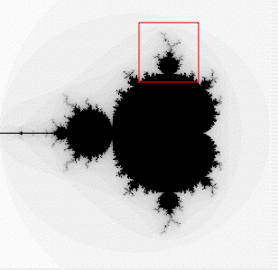
First, let’s talk about the structural physiology of lungs. Anyone who has ever dissected a mouse knows that the lungs collapse into a wet pink mess pretty much as soon as the organism dies. This is because mice have spongy lungs (as opposed to “simpler” bag-like lungs in many animals) like humans. This is also because lungs are mostly soft tissue with air-filled tubes running throughout, somewhat analogous to Jello with holes drilled through it (though I don’t know why anyone would drill holes in Jello, if it’s even drillable [I may need to empirically test this]). The trachea branches off into the main bronchi, which each branch into lobar bronchi. These then branch into tertiary bronchi and form bronchopulmonary segments separated by connective tissue (lobes, essentially). Inside the bronchopulmonary segments the bronchi are called segmental bronchi, which then divide into primary bronchioles before dividing further into terminal bronchioles. And because this isn’t yet enough (think about it: each stage of segmentation increases the surface area per given unit volume quite efficiently, like a Sierpinski triangle), the terminal bronchioles branch into respiratory bronchioles branch into alveolar ducts end in alveolar sacs that contain the alveoli. Alveoli are where gas exchange happens via the blood vessels that cover most of their surface area. As the airways branch further out, rigid supporting cartilage decreases and smooth muscle increases. This is important to note for later, as are the observations that each lung does not branch perfectly symmetrically.

Figure A: 2 ways to visualize the branching of the bronchi. Mandelbrot set visualization (top) and fractal tree (bottom).

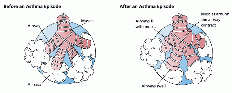
Second, let’s talk about the pathophysiology of asthma. The possible causes and/or triggers of asthma warrant an entire series of posts by themselves, so they won’t be discussed here. Asthma occurs when the lungs aren’t providing adequate respiratory exchange due to constricted air flow volume (also referred to as tidal volume) and the levels of oxygen in the blood begin to fall; asthma is generally treatable with drugs, this is what clinically differentiates asthma from COPD and/or some ARDS. Reduced tidal volume is primarily thought to be a direct consequence of increased smooth muscle activation. Smooth muscles can be triggered by a wide variety of extrinsic factors and the resulting asthma attack can range from mild wheezing to complete respiratory failure and death. Long-term pathophysiological manifestations include scarring of the lung airway epithelium, localized sustained inflammation, and a reduced baseline tidal capacity, which are thought to be due to recruitment of immune system effector cells into the underlying tissues (which in turn cyclically drives more inflammation).

Figure B: Like that (although this .png has been squished a bit).

Finally, then, to the paper. A computational model of a single terminal bronchiole that included the physics of positive airway pressure inside the lumen space, the tension of underlying parenchymal upon bronchioles, and the constriction caused by smooth muscle activation was integrated to simulate a whole lung by using a Mandelbrot-like structure that reflects the serial branching of airways. This model assumes constant tidal volume and rate and neglects gravity-dependent lung effects as would occur in the deeper lungs. It also ignores the effect of diaphragm muscles on lung function. Obviously, in real life we breathe harder when we’re winded and the air deep in our lungs has to contend with gravity more than air in our upper lungs, and we also use muscles to breathe, but simplifications are necessary to make modeling a sane endeavor.
And now for the metaphorical vehicle: Imagine you’re standing on a tightrope with someone else at some distance away from you, say, farther than you could throw a tuba. If you lose your balance, you’ll swing about to try to regain it, and doing so will move the rope you’re standing on, which will then result in the other person having to move to keep their balance. If you don’t quickly recover your balance and steady the rope, either you’ll fall or the other person will have to work harder to stay on the rope and continue to amplify your instability.

Figure C: Don't try this at home. Go somewhere more dangerous first.

Unfortunately, falling off the rope doesn’t really happen in asthma attacks (unless you think of that as a piece of lung shutting down). Sure, whole chunks of lung seize up and stop providing adequate ventilation, but this is patchy and even then those patches still contain smaller bits that still work as they’re supposed to. But it is the amplification of instability that leads to the progressive and terrifyingly fast clamping shut of airways in an asthma attack. Section A shuts down, so Sections B, C, and D will have to work harder to compensate. Compensation will involve greater intrabronchial airway pressure, which the normally functioning lung interprets as bad and tries to compensate for in the opposite direction by activating smooth muscle, so smooth muscle activation and airway constriction can spread rapidly from Section A to B to C in sudden and catastrophic sequence even if only a little tiny bit of Section A started the whole thing.

The computational model used in this paper found that catastrophic shifts as described above don’t really happen unless some degree (~1%) of heterogeneity in lung response to constriction/smooth muscle activation is included. If the computational model was allowed to be completely symmetrical and if all lung tissue responded in exactly the same way, then nothing spectacular happened.

While I find the computational modeling stuff to be fascinatingly awesome, that’s not all they did. They also got mild to moderate asthmatic volunteers to inhale isotopic nitrogen gas inside of a PET machine during a chemically induced asthma attack to map where the gas was going (those were very brave asthmatics). In poorly functioning lung, iN2 would be trapped and remain visible on the PET while normally functioning, or lung hyper-functioning to compensate, lung would clear it quickly. These PET results validated their modeling outcomes in that asthmatic events are patchy in that neighboring bronchioles are more likely to become distressed than distant bronchioles, though not absolutely in that the patchiness of poorly ventilated lung was itself patchy (mirroring computationally induced heterogeneity). Furthermore, these findings were clinically relevant because only normal- or hyper-functioning lung remained open during the asthmatic event, which suggests that inhaled anti-inflammatories may be being delivered primarily to the lung epithelium that needs it least.

Research Cited:
Jose G. Venegas, Tilo Winkler, Guido Musch, Marcos F. Vidal Melo, Dominick Layfield, Nora Tgavalekos, Alan J. Fischman, Ronald J. Callahan, Giacomo Bellani, R. Scott Harris (2005). Self-organized patchiness in asthma as a prelude to catastrophic shifts Nature, 434 (7034), 777-782 DOI: 10.1038/nature03490

*From my perspective as someone who doesn't actually do atopy research...yet, anyway. Therefore, if I am flatulating ignorance and posing it as wisdom, feel free to correct my mistakes in comments.

NOTE: This is my first post for the Cyber Journal Club that Science Bear is organizing. R.E.S.E.A.R.C.H.E.R.S are also participating. My goal is to post every Wednesday and apparently Science Bear is going to be posting 2 Wednesdays a month. Feel free to join in by commenting or posting your own (we'll ping you).