Showing posts with label research blogging. Show all posts

24 July, 2009

Optimizing Algorithms for Brain-Machine Interfaces

ResearchBlogging.orgImagine waking up trapped in a prison of your own flesh, blinking awake in the dull glow of a softly bleached hospital room. Your arms and legs are unresponsive to the will to move them, to the simple desire to reach up and scratch the itchiness of morphine from your eyes. Nothing happens, nothing responds, nothing moves, nothing feels. You are an immobile head trapped on an unresponsive body, and no matter how loudly you scream against the walls of your confinement from inside your head, nothing happens.

Luckily many quadriplegics retain the ability of speech and independent respiration. However, their quality of life, and that of the more unfortunate patients who are fully lucid but cannot communicate with the outside world by anything more than eye-blinking Morse code, remains severely compromised due to poor rehabilitation prospects and dependency upon caretakers. Perhaps one day we'll be able to repair injured nerves and restore connectivity with the rest of the body, but for now we're beginning to work out how to directly interface the brain with mechanical actuators. The goal is to develop an interface through which a paralyzed patient could control a machine that would augment their standard of living using nothing more than their mind and some technology.

Nerves and computers and both conveniently electrical. The problem is that they each operate on very different electrical schema. With computers we more or less know which transistor is storing which bit of information and have discrete units of conductors, resistors, capacitors and such. But it is very difficult to isolate the behavior of a single neuron in situ, so instead we use mass behavior of relatively large numbers of neurons to try to approximate functional firing chains. To translate the chaotic, non-discrete signal from neurons into discrete signals for machines, we need an algorithm. And to develop that algorithm, we need electrode-implanted animal models (unrestrained monkeys in this case) and scientists who can do math.

Li and Nicolesis et al have developed a novel modification of pre-existing brain-machine interface algorithms in "Unscented Kalman Filter for Brain-Machine Interfaces". Previous brain-machine interface algorithms were either linear approximations or non-linear particle filters. Linear filters (such as the Wiener and standard Kalman filters) didn't do as well approximating the behavior of neurons as particle filters, but they were much faster and computationally cheap enough that they could be used in real time. In general, particle filters (such as SSPPF) were very good at modeling the behavioral input of neurons, but they were so cumbersome that each iterative point required computing time that put it outside the range of useful real-time applications. Li and Nicolesis et al have modified a Kalman filter such that it uses a nonlinear tuning method (a quadratic model instead) and adds historical regression with multiple time offsets to help predict future behavior patterns. The quadratic tuning model integrates previous neural activity to arm movement models (the cosine tuning model, tuning to speed, tuning to distance of reach) into one cohesive and flexible equation. The historical regression with time offsets gives the model a short memory that allows it to quickly predict possible future states of neural activity to arm movement correlates and optimize them based on current neural activity input.

Implementing the quadratic neural tuning model greatly improved the accuracy of predicted neuron firing behavior, which means that the algorithm was better at interpreting the noisy signals from the neurons. At the same time, the historical regression made brain-activity-guided movement of the prosthetic hand* smoother and also helped to tune the monkeys' training on the algorithm by operating as a sort of continuous optimization that kicked out inefficient processes to improve overall performance. This quadratic model also significantly improved the reconstruction of the monkeys' desired hand movements during real-time tests in comparison to standard Kalman and Wiener filters.

In effect, this new algorithm construction allows for more accurate control of a prosthesis, with low tuning demands and progressive learning of efficient movement correlates. Ideally, this will one day allow paralyzed patients to accurately control mouse cursors (better than existing technology, anyway) or robotic prostheses that will greatly improve their quality of life. This technology may still be crude, but it is progressing rapidly and with great potential. I, for one, welcome the prospect of an auxiliary robotic arm. It would be great for my benchwork productivity.

*In this case, a cursor on a computer screen.

Li, Z., O'Doherty, J., Hanson, T., Lebedev, M., Henriquez, C., & Nicolelis, M. (2009). Unscented Kalman Filter for Brain-Machine Interfaces PLoS ONE, 4 (7) DOI: 10.1371/journal.pone.0006243

___________________________________________________


This is my entry in the Scientists' Duel that Hermitage and I are fighting for the title of Most Nefarious. Her entry is here. You, dear reader, will decide who wins. As of 12:00AM, 7/24/09, you have 72h to vote. You get 100 points to divide between Hermitage and I as you see fit. Report your scoring in comments. At the end of 72h we will tally up the points and determine the winner.

13 July, 2009

On Apoptosis in Development

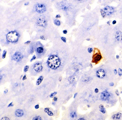
ResearchBlogging.orgApoptosis means doom for an individual cell. As such we tend to automatically assume that apoptosis is a Bad Thing, but in reality apoptosis often is quite necessary for normal physiological function at the organism level. In order for our bodies to maintain the homeostasis that defines so many of our cellular processes, we have to sacrifice some cells. As it turns out, we actually wind up sacrificing enormous numbers of cells every day. Worn out red blood cells, dangerously self-reactive lymphocytes, individual columnar epithelial cells and others. These processes are tightly regulated, so much so that most cell types actually require biochemical signals from neighboring cells, tissues, or even distant organs just to tell them to keep living. The anti-apoptotic survival signals can fall below a threshhold value and/or be overridden by pro-apoptotic stimuli, which normally results in swift induction of the apoptotic program. When individual cells develop mutations that deafen them to these signals, they become dangerous proliferation-happy pre-cancerous cells more interested in their own survival than that of their constituent organism.

Figure A: TUNEL histochemical staining in murine liver, brown cell is apoptotic.

The apoptotic program ultimately results in highly oxidative and degradative enzymes (such as proteases) hidden away in the mitochondria being released into the cytoplasm to wreak havoc. Usually the raw material of a dying cell is tidily absorbed by its neighbors to be recycled. I've always imagined mitochondria as pulsing with a low, gentle buzz in normal cellular physiology with occassional metallic pings as statistical flucuations in the net free energy of electrons falling down the electron transport chain is captured in ATP. Following this, I think the sound of caspase-8 et al slicing open the mitochondria would be like the initial panicked braking shriek of a train loaded with Furbies who are quickly drowned out in the self-amplifying roar like a tornado grinding through a gravel pit as the apoptotic effector enzymes set to work dissolving the cell from within.

Apoptosis is absolutely essential not just to adult homeostasis, but also to normal ontogeny. Without apoptosis organs would fail to separate, fingers would remain stuck together, and many other things would go very, very wrong. There are 2 families of intracellular proteins that battle to determine whether or not a cell will become apoptotic: the (generally) pro-apoptotic Bcl-2 family and the (generally) anti-apoptotic IAP family. Conveniently, IAP stands for Inhibitor of Apoptosis Protein. A recent review by Dr. O'Riordan et al discussed the diverse and essential roles for IAP proteins in normal tissue development across a wide range of model organisms. From ablated organ development in the absence of Diap1 in Drosophila larvae to stunted hematopoeitic developmental repertoire in the abscence of Survivin in mice, IAPs seem to be evolutionarily conserved signal transducers that integrate diverse extracellular signals into a coherent cellular action. Developmentally, the IAP proteins seem to be involved in everything from proper vascularization to chromosome stability, although it is important to note that direct modulation of apoptosis in developmental processes has only been established in invertebrates. Lack of any one of several IAPs in higher chordates has not been directly linked to developmental apoptosis, but several abnormal embryonic phenotypes and attenuated adult functional capacities have been demonstrated.

IAPs are grouped by the prescence of BIRs (baculovirus IAP repeats) and many also have RING domains. Both motifs have been found to have zinc-finger conformations and the interaction of different sections of adjacent BIR motifs in some proteins, such as direct inhibition of pro-apoptotic caspases-3 and -7 by BIR2 of XIAP (X-linked inhibitor of apoptosis protein), has been found to modulate a number of diverse effects. These diverse effects are potentiated by the ubiquitin ligase activity that some RING domains have demonstrated. IAPs help the organism balance necessary apoptosis and unnecessary apoptosis, and because apoptosis is required for the homeostasis of most tissues the IAP family has been evolutionarily conserved and biochemically diversified. IAPs remain an active and engaging area of research that holds great promise in the treatment of pathologies from cancer to intracellular bacterial infections and underscore how a little sacrifice for the team by one cell can make a massive impact on the constituent organism's overall fitness.

IAPs have also been found to modulate innate immunity, which will be discussed in another post.

ORIORDAN, M., BAULER, L., SCOTT, F., & DUCKETT, C. (2008). Inhibitor of Apoptosis Proteins in Eukaryotic Evolution and Development: A Model of Thematic Conservation Developmental Cell, 15 (4), 497-508 DOI: 10.1016/j.devcel.2008.09.012

Additional Source: Molecular Biology of the Cell; Alberts et al; 4th ed.; pages 1010-1014

01 July, 2009

Llamas Against Breast Cancer

ResearchBlogging.orgWe all knew llamas were kind of weird. They're fluffy. They're smelly. They spit. They're like Sanrio (the Hello Kitty company) tried to make over a camel. But that's not all. Llamas are also immunologically strange. Whereas most all other organisms with a humoral immune system produce large, multi-domain antibodies with several distinct genetic and structural motifs, llamas instead make nanobodies. Nanobodies are, basically, tiny little antibodies. Normal antibodies contain 2 heavy chains and 2 light chains, which each have V (variable, where the epitope binds) and J (joining) regions; and the heavy chains also have C (constant, these make up the Fc fragment) regions. All of those chains are bound together by disulfide bonds and the resulting antibody typically has 2 binding sites, each at the tip of the Y shape. Nanobodies dispense with all of that and only retain a functional binding site with a single variable light chain domain. As a result, nanobodies are much much smaller and can access and bind to sequestered epitopes or complex 3D epitopes that may otherwise be hidden inside a molecular cleft.


Figure A: Structual phylogenetic picture, much like a family picture. This is only an approximation as Good Images are copyrighted and Photoshopping ribbon-style molecules is difficult.

The point of this study is not that nanobodies are weird and cool. Instead, Alvarez-Rueda et al harnessed the complex 3D structural variability of nanobodies to mimic the immunogenic structure of HER2. HER2 is a surface protein normally only expressed in fetal development that is reexpressed in 20-40% of breast cancers and 30% of ovarian cancers. It is a member of the epidermal growth factor (EGF) family and is suspected to help cancerous cells proliferate more rapidly and aggressively. Tumor expression of HER2 correlates strongly with increased metastatisis and decreased survival. We've known about HER2 for a while now and it has been a target of intense research. There are now genetic tests available for HER2 alleles that correlate with increased morbidity from breast cancer. There was also a passive immunotherapeutic treatment against HER2 approved for use in combination with chemotherapy in 1998 called Trastuzumab (marketed as Herceptin(R) and manufactured by Roche). Trastumuzab is a humanized antibody therapy that targets HER2 directly; it is thought that it mimics the natural humoral immune response to HER2, which is observed to slow down tumor growth in early tumors but unfortunately sometimes fails to stop it. The primary problem with Trastuzumab is that it must be repeatedly administered over the course of cancer treatment to have any effect. While Trastumuzab is an invaluable tool in the fight against these cancers, it has long been recognized that inducing a robust host immune response would help to combat the tumor itself, and subsequent induction of a host immune memory against HER2 would help to prevent relapse of the cancer.

The best way to do this is with a vaccine.

Simply injecting HER2 with an adjuvant could produce a strong immune response, but the HER2 itself could make the cancer worse meanwhile. The ideal vaccine would be a molecule that mimics the structure of HER2 closely enough to induce cross-reactive immunity but that doesn't have the biological activity of HER2.

Enter the llama and its nanobodies.

Alvarez-Rueda et al injected Trastuzumab into a llama and the llama kindly produced nanobodies. Because Trastuzumab is an antibody against HER2, the llama's immune system produced a molecule against it that is somewhat structurally similar to HER2. When this nanobody molecule was isolated and expressed via transgenic clone library, it was found to strongly bind both Trastuzumab as well as isolated human anti-HER2 antibodies. So they then took the nanobody (called 1HE) and injected it into mice (along with Freund's adjuvant). As expected, the mice produced antibodies against the nanobody. These antibodies then, in turn, bound strongly to both 1HE and the HER2 protein. This strongly implies that immunization of a human with the 1HE nanobody and adjuvant would induce a strong anti-HER2 antibody response, effectively immunizing them against HER2-expressing breast or ovarian cancers or arresting the growth of existing tumors as part of chemotherapy*. Additionally, the polyclonal antibodies produced by the mice in response to the nanobody were found to inhibit growth of HER2-expressing carcinoma cell lines. The data have not yet been validated in vivo.

Either way, this is a cool advance in the fight against breast cancer, and I sincerely hope that something therapeutically useful in humans will soon come out of this. Thank you, llamas.

*This is technically known as an anti-idiopathic vaccine.

Alvarez-Rueda, N., Ladjemi, M., Béhar, G., Corgnac, S., Pugnière, M., Roquet, F., Bascoul-Mollevi, C., Baty, D., Pèlegrin, A., & Navarro-Teulon, I. (2009). A llama single domain anti-idiotypic antibody mimicking HER2 as a vaccine: Immunogenicity and efficacy Vaccine DOI: 10.1016/j.vaccine.2009.05.067

27 June, 2009

Silky Muscles

ResearchBlogging.orgYou're running through the cool woods on a hot day, barefoot as dead leaves rustle underfoot and the cold flint tickles beneath. The green leaves and kudzu blur past as you dodge beaming shafts of sunlight and the hot ground they illuminate. You scan the earth ahead for sinkholes and patches of poison ivy, but still, the chilled, humid air coiled around the trees flowing in your ears feels joyous in comparison to the sauna of the open field. You dart between two trees, then suddenly stop and gyrate wildly, windmilling arms about your face as you splutter and ick; swiping instinctually at your face to pull away the clinging threads of a spider web.

To many of us, spiders are mostly nuisances, either by dangling from a single invisible thread in the most inconvenient places or by stumbling into webs and getting their sticky strands stuck in our eyebrows such that we look like a surprised Gandalf. However, a recent publication suggests that spider silk, the material they spin webs and drag lines out of, may turn out to be much more useful that we previously thought.

Compared to contractile biological muscles, mechanical rotary motors are rather inefficient. Getting a micro-servo to function correctly in a robot arm is a difficult art of soldering and fine-tuning. In prosthetic limbs, robots, and industrial applications, there is a current need for a small, reliable, lightweight, and dependable actuator. It turns out that when spider silk is exposed to alternating extremes of ambient humidity*, it contracts much like a biological muscle and does so repeatedly. Many biological fibers (cotton, wool, etc.) can also contract in high humidity, but they can only do so once before becoming inert. This occurs because different fibers are composed of repetitive hydrophilic materials that suck up water and collapse into lower net energetic states when the water is available, as it is during humid conditions.

The really cool part of this research was the force generated by the spider silk. On a basis of equivalent mass, spider silk was found to be capable of doing 500X the work of a human biological muscle. Agnarsson et al calculated that, based on their scaling experiments with combining individual silk fibers, a 2cm diameter strand of spider silk would be capable of lifting 2tons of mass! Similar, though weaker, effects were observed in silkworm silk (which is already commercially availble).

The caveat to this, because there's always a caveat, is the degree to which the spider silk contracts. Human muscle is capable of elastic modulus (how much it can bunch up without breaking) of 30-40%, while spider sillk was found to be capable of a modulus of only ~2%. This is considerably less useful, but still cool. The researchers noted that this was all done in one particular species of spider, Nephila clavipes, and that the silk of other spider species may turn out to have more useful modulus while preserving greater scaling strength and simple humidity switch.

Let's hope that these tests are done and something found, because robots everywhere are itching for a change.

*Steps of 10% differences. Contraction was found to be irreversible after exposure to <70%>

Agnarsson, I., Dhinojwala, A., Sahni, V., & Blackledge, T. (2009). Spider silk as a novel high performance biomimetic muscle driven by humidity Journal of Experimental Biology, 212 (13), 1990-1994 DOI: 10.1242/jeb.028282

27 May, 2009

Your Microbiome and You

ResearchBlogging.orgYou are never alone. Not even when you might want to be. Tucked away within the ~100m2 of your bowels are ~1014 (there are ~1013 somatic and germinal cells in the human body) of your closest friends, collectively termed The Microbiota. They eat, spawn, conjugate, die, poop, fight, and secrete right there inside of you, unseen and mostly unthought of except when something is wrong. This system, the remarkably homeostatic mammalian gut, forms what is perhaps the densest and most complex microbial ecology on this planet.

These teeming microbes are not mere freeloaders living off of your access at their own convenience, they are true symbionts. In exchange for a warm, wet home and nutritional supply, they break down starches for us, metabolize complex molecules, and synthesize some key compounds, such as Vitamin K. It has been found that gnotobiotic, or germ-free, animal models require ~30% more calories to develop normally without a microbiota to help them out. In humans that have been on a broad-spectrum antibiotics, hardier inhabitants (such as Clostridium difficile) can bloom when all of their more sensitive neighbors (such as Bacteroides spp. and Bifidobacterium spp.) are killed off, which causes very unpleasant colitis and diarrhea, that can then be cured by a transplant of fresh microbiota from a healthy individual (colloquially referred to as "poop soup"). Microbiome transplants can also transfer physiological characteristics from one individual to another. For example, the microbiomes of obese individuals have been found to have reduced numbers of Bacteroidales spp., and transfer of these microbiota via poop soup into germ-free mice resulted in obese mice, theoretically because these microbiota were more efficient at releasing calories from food.

Microbes exist, or can exist, in virtually every segment of the gastrointestinal tract from mouth to anus. In the mouth, a variety of Actinomyces spp. are associated with the formation of plaque. In the forbidding and harsh environment of the stomach, only Helicobacter pylori can thrive (it does so by hiding among the mucous lining the stomach and modulating the host immune response) and it has been found to directly cause stomach ulcers and has been further implicated in the formation of gastric cancers (it's the only organism classified as a BSL 2+ carcinogen). The proximal portion of the small bowel is relatively sparsely colonized at ~104-105 microorganisms/ml lumenal contents, which contrasts sharply with the densely colonized colon (~1010-1012 microbes/ml contents).

In the human and other mammals, diverse and distinct microbial ecologies also exist in the sinuses, ears, genitourinary tract (largely Lactobacillus spp. in the vagina; the bladder is generally only colonized in disease states [long-term catherization and/or pyelonephritis] by uropathogenic Escherichia coli, Proteus mirabalis, et al), and on the skin as a whole (mostly Staphylococcus spp.). These others will, however, be excluded from the present discussion.

However, what's very puzzling about all of this is: how does the mammalian immune system manage to differentiate from the massive basal antigenic signals coming from the microbiome from pathogenic antigens? In other words, why isn't the immune system raging against the huge number of microbial signals in the gut?

One of the exquisitely elegant features of normal gut physiology is that gut-associated lymphatic tissues (GALTs) mediate fine-tuned hyporesponsiveness to commensal microbiota while remaining responsive to pathogenic microbes. This flies directly in the face of most immunology, which holds that microbial antigens will always provoke a stimulatory response when ligated to TLRs, CLRs, or NODs (conserved receptors of the immune system that bind conserved molecular patterns associated with pathogens). In vitro data support this. Physiology doesn't.

Physiologically, the germ-free mouse is weird. A germ-free animal is one that has been reared in an environment completely free of all microbes, fungi, and exogenous viruses and as such they have no native intestinal microbiota. Not only do they require more calories and vitamin supplementation, but they also tend to accumulate undigested fibrotic material in their ceca, which predisposes them to gut twists and bloat. Additionally, they feature underdeveloped Peyer's patches (distinct GALT sites on the gastric mucosa), altered CD4+ T-cell and IgA-producing B-cell population profiles, and the follicles in the spleen and lymph nodes where T- and B-cells mature are poorly formed. All of these abnormalities can be rescued by adding back microbial signals such as LPS, even without the microbes themselves. Due to these alterations, it is becoming accepted that the microbiome plays a crucial role in the normal development of the immune system. But to reconcile this with the dogma of microbial signal + PRR ---> inflammatory immune reaction is somewhat difficult, or at the very least complex.

Immune cells that reside in the lamina propria underneath the gastric epithelium generally show signs of recent activation and a particular subset of dendritic cells (CX3CR1+) has been found to extend dendritic processes up through the tight junctions binding gastric columnar epithelial cells together to directly sample the lumenal contents. M cells that cap the Peyer's patches have been found to shuttle lumenal contents, and any antigens contained therein, to the dendritic cells and lymphocytes underneath. These pathways of antigen exposure are thought to be involved in the induction of immunological tolerance to microbiotal antigens, which could explain why the immune system does not attack the commensal microbiota. However, it does not explain how pathogen antigens processed by the same pathways are recognized as pathogenic and stimulate the immune system to attack.

Recent evidence strongly suggests that the intestinal epithelium itself is responsible for the differentiation of nonpathogenic microbiota from pathogens. Canonically, the intestinal epithelium is thought of as a simple barrier that is involved in the absorption and transcytosis of metabolites and nutrients. But it seems that it is much more involved that we had previously believed.

It turns out that intestinal epithelial cells (IECs) express TLRs and directly modulate the composition of the microbiome itself as well as the responsiveness of immune cells. This ranges from TLR expression on Paneth cells in the small intestine that secrete potent antimicrobial molecules (RegIIIg) when ligated [Dr. Lora Hooper, in seminar given 11/19/08] to actual expression of MHCII and direct antigen presentation. It was previously believed that MHCII expression was restricted to antigen presentation by dendritic cells.

When investigators deleted TLR4, NOD1, or MyD88 (an adapter protein involved in many TLR-mediated NF-kB inflammatory pathways) in murine IECs they found that the mice were more susceptible to bacterial infections, which implies that the TLR signalling on the IECs is essentially to the development of normal protective immunity. A second feature of this is that IEC TLRs and NODs are located intracellularly, instead of on the cell surface as in immune cells, which means that they'd only be ligated and activated when an invasive pathogenic microbe breaks into the IECs themselves (e.g., Salmonella typhimurium, Vibrio cholerae) as opposed to the more peaceful commensals. It may be that noninvasive gastrointestinal pathogens are recognized by the proteins that they shoot into IECs via Type IV secretions systems (e.g., Tir and Escherichia coli O157:H7) in the same manner.

The commensal microbiota is also at work on the IECs themselves, actively acting against IEC-mediated inflammation. Bacteroides thetaiotaomicron has been found to induce the PPARg anti-inflammatory (acts by increasing cytoplasmic shuttling of pro-inflammatory NF-kB away from the nucleus) mechanism in vitro. Commensal-derived metabolites such as butyrate (a short-chain fatty acid) have been found to inhibit expression of pro-inflammatory cytokines and increase expression of anti-inflammatory cytokines in IECs.

It is now thought that IECs regulate dendritic cell function through secretion of thymic stromal lymphopoietin (TSLP) and modulate T-cell activity through expression of MHCII in the abscence of costimulatory molecules. TLSP acts directly on dendritic cells and inhibits their production of pro-inflammatory cytokines (such as IL-12), which in turn promotes dendritic-cell-mediated activation of regulatory T-cells. TSLP is also implicated in skewing the immune response to a TH2-type T-cell response, which is implicated in both response to metazoan parasites and pulmonary atopy. If naive T-cells are being exposed to MHCII on IECs without co-stimulatory molecules, then the T-cells will either kill themselves off (anergy) or mature into tolerogenic T-cells that limit the immune response to those given antigens. This, combined with widespread TGFb secretion by IECs, directly indicates an active role for IECs in promoting immune system hyporesponsiveness to the antigens present in the gastrointestinal system. Without this direct suppression of active, inflammatory immune responses, the immune system would be in a continual inflammation state due to not knowing what to do with a safe commensal antigen vs. a dangerous pathogenic antigen. Indeed, emerging research indicates that dysregulation of this process may underlie the pathophysiologies of inflammatory bowel disease and Crohn's disease.

It'll be interesting to see what's found next.

Artis, D. (2008). Epithelial-cell recognition of commensal bacteria and maintenance of immune homeostasis in the gut Nature Reviews Immunology, 8 (6), 411-420 DOI: 10.1038/nri2316

15 May, 2009

Breast Milk Transfer of Antigens Establishes Anti-Allergenic Tolerance to Those Antigens

ResearchBlogging.orgMilk isn't just milk. The pasteurized cow milk that we can purchase in the grocery store has been cleaned, processed, and in many cases chemically scrubbed of fat. Unique among mammals, we Western humans stubbornly persist in our consumption of dairy products well into adulthood regardless of whether or not our guts like it. But the cow milk in the store is very different from the fresh milk humans nurse their newborns with. Fresh human milk contains growth factors, vitamins, an astounding density of calories, IgA antibodies, and also antigen. Disregarding the controversial and impassioned debate surrounding breast milk vs. infant formula, research has found that the immunological molecules secreted in breast milk are important for the developing immune system of the infant.

Verhasselt and Julia et al have demonstrated that antigen secreted in a mother's breast milk significantly impacts the later development of allergic asthma in her infants. In effect, this is immunological programming.

They used an elegant experimental system wherein they took nursing dams (mouse mother) and exposed them to antigens (OVA) without their pups, then placed them back with their pups to nurse. Later on when the pups had reached adulthood, they sensitized the mice per the immunology canon and tested their allergic asthmatic (hereafter refered to as atopic) response. In mice breastfed by OVA-exposed dams it was found that, in comparison to those breastfed by non-exposed dams, airway hyperreactivity, pulmonary eosinophilia, cellular infiltrate, and mucus deposition were all decreased towards mice not challenged with OVA (normal, negative controls). Moreover, the classical TH2 cytokines (IL-4, IL-5, IL-10, and IL-13) that have been associated widely with atopic responses were decreased in OVA-breastfed mice, as were the frequencies of the lung CD4+ T-cells that secrete them. Overall, this points to OVA-breastfed mice having a significantly weakened allergenic response to an allergen, or put another way, these mice tolerated the prescence of the allergen much better.

As an aside, OVA is an abbreviation for ovalbumin, which is a key molecule in eggs and is a very sticky molecule. In every study of allergy or asthma that I can remember reading, OVA was used to sensitize mice and produce an allergic response to it. This is done by injecting a solution of OVA into the peritoneal cavity of the mice, waiting 2 weeks to let OVA-specific CD4+ T-cells develop, and then challenging the mice by squirting an OVA aerosol up their noses. This model has proven to reliably produce a strong and specific allergenic response.

But how did the authors determine that it was secreted antigen itself that was inducing the tolerance?

To address this, they took normal, wild-type C57 mouse pups and gave them to lactating uMT and RAG-2 transgenic dams*. Both these transgenic strains of mice are completely unable to mount an adaptive immune response to anything; they do not and cannot make antibodies. When these mice were exposed to OVA and then nursed the wild-type pups, the same effects of the OVA-breastfeeding inducing allergen tolerance were observed. This was also replicated with Balb/c mice (which is important because the C57 strain is known to skew towards a TH1 response phenotype while Balb/c skews more to TH2, which is better characterized in the pathophysiology of atopy).

But then Verhasselt and Julia took a look at some important and specific immunoregulatory molecules: TGFb and IL-10. Both of these molecules are broad suppresors of inflammatory immune responses regardless of that response's cellular phenotype. IL-10 transgenic dams did not alter observed results when exposed to OVA, but TGFb knockdown dams did in that the pups they nursed were reactive to OVA. This strongly suggests that TGFb must accompany antigen in the breast milk in order for the infants' immune systems to recognize that antigen as a harmless allergen and, in effect, program itself not to react against it. Luckily for human mothers, though, breast milk already contains TGFb.

At a wider level, this suggests that mothers who are exposed to many allergens will pass on tolerance to those allergens to any breast-feeding infants they may have. Perhaps this, coupled with the hygiene hypothesis, is a call for more moms to teach their infants how to make mud pies even earlier.

*I can't help but wonder how many times they did this and found all the pups had been killed off by their adoptive mothers, as this is what stressed out rodent dams tend to do.

Verhasselt, V., Milcent, V., Cazareth, J., Kanda, A., Fleury, S., Dombrowicz, D., Glaichenhaus, N., & Julia, V. (2008). Breast milk–mediated transfer of an antigen induces tolerance and protection from allergic asthma Nature Medicine, 14 (2), 170-175 DOI: 10.1038/nm1718

20 April, 2009

Developmental Effects of Ambient Air Pollution

ResearchBlogging.orgFirst off, props to Dr. Isis, who in the discussion of her Cairo haze post referenced the work of L. Calderón-Garcidueñas, which set me off into the literature. What I have initially found was sobering.

Air isn't something we often think about. We take it entirely for granted. We complain about how hot or humid it is, but we usually don't have to think about what in that air might be making us, or our children, sick. Those of us fortunate enough to live in Western, industrialized countries really are well-off in that we don't ever really have to pit our own health against the simple act of breathing. Other areas in the world aren't so fortunate.

Dr. Calderón-Garcidueñas' work focuses on the effects of airborne pollutants and particulate matter on the developing lungs of children in Mexico City, Mexico. But her work is actually relevant to anywhere that the air isn't shiny clean.

By radiograph, her team found that children in Mexico City with lifelong exposure to the polluted air had significantly increased rates of lung hyperinflation when compared to age-matched controls from a much less polluted area (Tuxpam, Ver [I'm guessing this is somewhere else in Mexico?]). Lung hyperinflation by itself in one person doesn't indicate a lot, it just means that there is more air in the small alveoli of the lung so it appears larger on a radiograph. This could mean asthma, emphysema, or even lung cancer, but could also just mean the patient was breathing hard. But when there is such a significant (p=0.0004) between groups it becomes indicative of pollution-induced lung dysfunction.

This is in addition to previous work demonstrating that children in this severly polluted area of Mexico City had altered nasal apparatus such that the mucociliary clearing/filter mechanism wasn't working so well as it should be. This is troubling because the loss of the nasal ciliary filter leads to an even greater dose of reactive gasses or particulate matters getting past it into the lower lungs, thus exacerbating the initial problem.

Referenced experiments in this paper showed even more going on. Particulate matter (PM) less than 5um in aerodynamic diameter was found to disproportionately wind up in the alveolar sacks of the lung while PM greater than 10um was found to locate to the proximal bronchioles. PM doesn't seem to affect lung epithelia directly, but it does activate alveolar macrophages to produce inflammatory cytokines (IL-6 and TNF) that utlimately will attract additional inflammatory infiltrate and further damage lung tissues. On the other hand, reactive gasses were found, in vitro and in animal models, to elicit secretion of IL-6, IL-8 (both inflammatory), and fibronectin (involved in repair if tissue damage) in lung epithelial cells.

Together, these 2 overlapping respsonses indicate the air pollution is driving a repetitive damage-repair cycle in lung tissues. Pollution damages lung tissue and activates the immune system to produce and inflammatory response. So the lung tissue tries to repair itself in an inflammed environment, which can lead to scar-like tissue regeneration. The altered tissue regeneration (as in the loss of the nasal cilia above) can lead to lung tissue more vulnerable to subsequent pollution injury. Over time, this kind of cycle leads to long-term tissue remodeling such as increased constrictive reactivity of existing smooth muscle (a hallmark of acute asthmatic responses), increased smooth muscle metaplasia (chronic asthma), eosinophilia (asthma and allergy reactivity), and even destruction of the walls dividing alveoli (a hallmark of emphysema). And what's saddest about this is that moving animal models of exposure to clean air did not completely reverse the exposure-related pathologies, which means that the lung damage will be a life-long legacy of any children that grew up in it.

So take a deep breath, and be thankful it's mostly clean.

Calderón-Garcidue˜nas, L. (2000). Respiratory tract pathology and cytokine imbalance in clinically healthy children chronically and sequentially exposed to air pollutants Medical Hypotheses, 55 (5), 373-378 DOI: 10.1054/mehy.2000.1070

(P.S. - Whomever came to this blog searching for "burning bronchioles so they won't constrict", please don't. I can think of few worse ways to die than by one literally drowning in necrotic, burnt lung tissue while gasping painfully for whatever air they can still get.)

10 April, 2009

No Dendritic Cells --> Unchecked CD4+ T-Cell Proliferation --> Autoimmunity --> DEATH

ResearchBlogging.orgDendritic cells (DCs) are the extremely important interface cell type between the innate and adaptive immune systems. Without them, the adaptive immune system has a very hard time getting started and the organism has a much more difficult time fighting off pathogens. But DCs don't just save our lives when we're sick, they also save us from ourselves when we're not. It has recently been found that constitutive knockout of dendritic cells leads to the development of spontaneous fatal autoimmunity.

[Context/background in previous posts on dendritic cells, T-cells, asthma T-cells, and multiple sclerosis autoimmunity.]

As is the case in many immune system pathologies, this comes back to T-cells. In a normal thymus, T-cells are continually proliferating in huge numbers. Each T-cell undergoes Rag-mediated receptor specificity recombination and randomization while it is forming its receptor. This gives rise to T-cells with receptors specific to a massively diverse array of potential antigens and is at the core of how the adaptive immune system recognizes pathogen peptides and fights back against them specifically. However, most of the T-cells that get made are killed off summarily for having receptors that react against self-antigens (meaning peptides native to the organism; called autoreactivity). Although the thymic epithelium is also involved in screening potential T-cells for autoreactivity, DCs are the primary mechanism by which these potentially harmful T-cells are weeded out before they can escape from the thymus differentiated and initiate an autoimmune response. There is even a specialized subset of thymic DCs that express diverse self-antigens to help weed the T-cells reacting against them out, and systemic plasmacytoid and myeloid DCs often circulate through the thymus or lymph nodes bearing chunks of apoptotic self-cells.

This group found a way to constitutively delete DCs. Without the DCs, the CD4+ T-cells of the mice went wild and proliferated massively because they weren't being tested and usually killed anymore (~10-fold more IFNg- and IL-17A-producing CD4+ T-cells in DC- than DC+ mice). This allowed self-reactive T-cells to mature and get out into the circulation, where they infiltrated various tissues and caused massive inflammation. Many of the mice used here died after the autoimmune response had developed. It should be noted that the DC deletion wasn't absolute, IFN-producing DCs were unaffected by their construct and B-cells and some macrophages have been shown to be able to prime T-cells as well, which explains how the CD4+ T-cells were able to fully mature in DC- mice.

Not only did effector inflammatory CD4+ T-cells increase, but Treg cells increased in frequency also, they just didn't seem to be doing much. They seemed to see that effector inflammatory CD4+ T-cells were out of control and tried to proliferate to help curb this, but without DCs they couldn't actually make anything happen. This prompted a quick preliminary search through PubMed and Google Scholar and it looks like the IL-10 that gets produced by Tregs decreases DC maturation from monocytes and encourages macrophage formation instead. It also looks like immature pre-DCs exposed to IL-10 don't express as much CD4/8 or MHCI/II as fully differentiated DCs. This is interesting in that is strongly suggests Tregs don't act directly on inflammatory T-cells, but instead act through DC-mediated pathways.

The group also infected DC- mice with helminths to provoke an immune response and see what happens. Not only were DC- mice unable to clear the infection as DC+ mice were, but DC- also had reduced pulmonary eosinophilia and increased TH1 and TH17 type effector T-cell tissue infiltration (oddly, they didn't report on any TH2 type data, which would have been very interesting). This demonstrated that DCs were absolutely required to mount an effective immune response to infection, which apparently hadn't been definitively shown before (probably because it's hard to knock DCs out; see below).

However, even though DC- mice weren't able to clear a helminth infection, they were able to make antibodies. This defies classical immunology dogma because it is widely supported that T-cells go and activate B-cells to make antibodies after getting primed by DCs. Under some circumstances, e.g., this DC- one, T-cells can still get primed by B-cells or peripheral monocyte populations, which may include the IFN-producing DCs that didn't get knocked out.

To test for antibodies from DC- mice, they used tissue sections from rag-/- mice and immunofluorescently stained them for mouse IgG. Rag is the gene that controls the formation of unique T- and B-cell receptors, and without the rag gene the organism has absolutely no adaptive immune response* and makes no antibodies whatsoever, although they still do have innate immune effector cells. Therefore rag-/- mouse tissues will have no antibodies and no background staining, allowing detection of autoreactive antibodies from the DC- mice. The immunofluorescence staining showed that the targets of autoimmunity varied widely, with some mice autoreactive to nuclear components, others to lamina propria components, and still others to the epithelium itself. Why is this important? Antibodies are the adaptive immune system's signal to the innate immune system that something the innate immune system otherwise wouldn't see as bad is, in fact, bad and should be destroyed. So if something gets antibody bound to it, the innate immune effector cells (i.e., neutrophils, macrophages, eosinophils, even DCs) will recognize it as bad and chew on it. In the case of the DC- mice, the unregulated adaptive immune system wound up labeling the mouse's own tissue as bad and initiated inflammatory responses against it, sort of shooting itself in the foot.

In summary, DCs are important not only to fight off infection, but also to keep the adaptive inflammatory responses in check. Dysregulation or maldevelopment of DCs is therefore a potential target for remediation in autoimmune conditions such as inflammatory bowel disease, rheumatoid arthritis, and multiple sclerosis.

Genetics Addendum: The DC-knockout construct that this group made was really elegant and cool. I didn't mention it above because I wanted to stick to the cool big stuff, but this here is some awesome little stuff. Cre is a recombinase that recognizes floxP sequences. When there are 2 floxP sequences flanking a gene and when Cre gets expressed, it recognizes those floxP sequences and cuts out everything between. This system underpins 17 metric craploads of transgenic mouse studies.

Here, they built a construct where Cre was constitutively (this means always) expressed by a DC-specific promoter. They also stuck in a 3-layer sequence elsewhere on the construct. The outer layer of the sequence consisted of 2 complementary chunks of the sequence for diptheria toxin, the thin middle layer consisted of the floxP sequences, and the inner layer was an erythropoetin resistance gene. So without the Cre, the erythropoetin resistance gene disrupted the diptheria toxin gene. But when Cre got expressed in developing DCs in the bone marrow, the floxP sequences and the erythropoetin resistance gene got cut out and joined the diptheria toxin back into a functional sequence. So then diptheria toxin got made and killed the developing DCs before they could even get out of the bone marrow.

*This makes rag-/- mice popular for studying T-cell behavior. In these experiments, T-cells are taken from a rag+/+ mouse, manipulated according to experimental aims, and injected into rag-/- mice where their behavior and dynamics may be readily observed without native interference.


Ohnmacht, C., Pullner, A., King, S., Drexler, I., Meier, S., Brocker, T., & Voehringer, D. (2009). Constitutive ablation of dendritic cells breaks self-tolerance of CD4 T cells and results in spontaneous fatal autoimmunity Journal of Experimental Medicine, 206 (3), 549-559 DOI: 10.1084/jem.20082394

08 April, 2009

Muscle Hypertrophy

ResearchBlogging.orgHaving somewhat recently realized that I don't want to have the awesomely statuesque physique of a stick figure for the rest of my life, I began working out (before and after picture below). I still have no idea how to use most of the equipment in the gym and I'm not going to ask Pikkuveli (="little brother") how to use it just because he has ~130kg of muscle to my paltry ~60kg (we're also the same height: ~2.07m). But I am willing to ask where muscles come from.

So I hit up Current Opinion in Pharmacology and managed to learn something: muscles come from your liver.

Kinda weird, huh?

So here's how it more-or-less works:
1) ghrelin gets made by the fundus of your stomach
2) and binds to GHS-R in the arcuate nucleus of the hypothalamus, which then releases growth hormone in response
3) which travels to the liver and gets it to make up some insulin-like growth factor (IGF-1)
4) which then enters the systemic circulation and
5) acts in concert with local mechano growth factor (MGF) at the skeletal muscle to increase net protein synthesis and myotubule formation

But here's what's really cool and elegant about it: IGF-1 and MGF get made from the same mRNA transcript, just spliced up differently in different tissues.

Figure A: Electron micrograph showing a neuromuscular junction. M = muscle, T = axon terminus, arrow = junctional folds with basal lamina. Scale bar = 3um. Source: Wikimedia Commons.

MGF is produced by skeletal muscle tissue in response to mechanical stress (lifting heavy stuff) and cellular damage (the burn the day after working out) and acts in a paracrine and autocrine manner in and on satellite cells that hang out outside the membrane enveloping the muscle fiber. Satellite cells are mononucleated muscle stem cells. MGF tells them to proliferate and make more of themselves. But in order for this increase in satellite cells to translate into muscle growth, IGF-1 (IGF-1Ea, specifically) has to come along and get the proliferating satellite cells to cross the muscle fiber membrane and merge with each other to form a mature, multinucleated myotubule capable of contracting.

Conveniently enough, MGF levels are increased in skeletal muscle for ~2 days post-workout before tapering off. IGF-1Ea levels are increased for longer than that, so there is a sort of 2-phase muscle growth mechanism at work that smartly regulates itself. If MGF levels didn't taper off, satellite cells would keep proliferating and differentiating to myotubules and we'd drown in overgrowing buffness.

But, it's not quite so simple as that. Skeletal muscles are in a continual state of flux as they tear themselves apart and build themselves back up. The body can stash away amino acids in skeletal muscle or grab them back out as needed to help regulate serum pH. Muscle turnover also helps get rid of old, damaged myotubules and replace them with shiny, new, functionalier ones. Apparently this process takes about 2 weeks (I'm thinking maybe this is a minimum interval for exercise to maintain physique?) and heavily involves myostatin. Natural myostatin knockout mutants include Belgian blue cattle.

Figure B: A Belgian Blue bull (on the right).

Laboratory knockouts of myostatin in mice confirm that no myostatin to help muscle turnover along leads to increased muscle growth, but does not increase strength at all. This is thought to be due to increased accumulation of nonfunctional protein in muscle fibers.

Figure C: Toaster's physique before beginning exercise (left) and after (right).

So right about now my latest MGF pulse should be slowing and hopefully my IFG-1Ea is kicking in to get the new satellite cells that surely must be teeming in my shoulders and triceps fusing into functional new muscle. In the meantime, however, raising my elbows anywhere above shoulder level is rather more painful than I thought it might be. Maybe I shouldn't have done quite so many pull-ups, lat pull-downs, and tricep dips...


GOLDSPINK, G., WESSNER, B., & BACHL, N. (2008). Growth factors, muscle function and doping Current Opinion in Pharmacology, 8 (3), 352-357 DOI: 10.1016/j.coph.2008.02.002

01 April, 2009

Leptin, Ghrelin, Adiponectin, and Resistin: Fat and Hungry

ResearchBlogging.orgSo I recently realized that I don't know nearly enough about mammalian metabolism, or my own, really. Therefore, naturally, I turned to my best friends for answers: PubMed and Google Scholar. I learned some stuff. And now I'm going to learn you some stuff about the metabolic hormones.

We all know about insulin, even if we have no idea what it does. Basically, you eat stuff. Stuff has sugar. Stuff + stuff sugar gets absorbed across intestinal epithelium into your bloodstream. Serum levels of glucose rise and the beta cells in the islets in the pancreas are all like "Yo, I got this!", so they release insulin, which goes out into the bloodstream and tells the other cells, specifically those in hepatic and adipose tissues (and skeletal muscle for the picky), to get up and upregulate the surface proteins that take glucose out of the blood and do stuff with it. Like make glycogen or do some fatty acid reduction of glucose into triglycerides and stuff. Net result: insulin reduces bloodstream levels of glucose when they're high.

Not as many people know about glucagon. Glucagon is insulin's arch-nemesis. When bloodstream glucose is lower than it should be (when you haven't recently eaten), glucagon gets released and tells those same tissues that stored it to start releasing glucose back into the bloodstream so that tissues without much glucose/glycogen storage capacity (like your brain) can continue to eat and survive and stuff. Net result: glucagon increases bloodstream levels of glucose when they're low.

This is all well and good, but how do the stomach, brain, and fat communicate?

Enter Leptin, Adiponectin, Ghrelin, and Resistin.

Leptin makes you sated,
Ghrelin makes you hunger
Resistin's role is still debated
Adiponectin's from your blubber

Leptin is secreted by adipose tissue, which is just a polite way of saying that it's made by fat. The more fat present: the more baseline leptin gets secreted; although as the levels of leptin increase some fancy stuff starts to happen with its receptors, of which there are several isoforms. Leptin acts and peaks in concert with insulin to stimulate uptake of glucose and fat storage, but it also acts upon the hypothalamus to make you feel full after you've started eating. This is interesting because it suggests that feeling full has a lot more to do with blood glucose level's effect on the circulating leptin than actual stomach volume. Maybe this is why Snickers bars can sometimes be sorta filling. It is thought that in normal feeding patterns, leptin functions as a sensor of total body fat ratio and possibly as a link to the reproductive system as a switch on whether sufficient energy reserves are present for normal reproductive function.

Ghrelin is unique in that it is a hormone that comes from your tummy, directly from the fundus of the stomach. It binds GHS-R in the arcuate nucleus of the hypothalamus and has been shown to cause a release of growth hormone. Ghrelin makes you feel hungry and is associated with increased stomach motility and increased secretion of gastric juices (grumbling tummy).

Adiponectin is secreted by adipose tissue as well. Adiponectin increases insulin sensitivity in adipose tissue and also improves the glucose response. As BMI increases, adiponectin secretion decreases, thus acting kind of as a sensor for total body fat (although leptin kind of does that, too) but at the same time making fat less sensitive to insulin and thus more prone to diabetes.

Resistin is poorly understood in humans as it has been primarily characterized in rats. It is, however, associated with insulin resistance at increased concentrations and is secreted more at higher BMIs.

And this is just the tip of the iceberg for this subject. There're also orexins, neuropeptide Y, the effects of IGFs, PPARs, glucocorticoids, beta-adrenergic stuff and more. I know this post was a bit shallow on details and as such will probably offend real metabolic scientists, but it was a 15-page or so review, so I had to be be choosy.

Abstract:
BACKGROUND: Recent studies point to the adipose tissue as a highly active endocrine organ secreting a range of hormones. Leptin, ghrelin, adiponectin, and resistin are considered to take part in the regulation of energy metabolism. APPROACH: This review summarizes recent knowledge on leptin and its receptor and on ghrelin, adiponectin, and resistin, and emphasizes their roles in pathobiochemistry and clinical chemistry. CONTENT: Leptin, adiponectin, and resistin are produced by the adipose tissue. The protein leptin, a satiety hormone, regulates appetite and energy balance of the body. Adiponectin could suppress the development of atherosclerosis and liver fibrosis and might play a role as an antiinflammatory hormone. Increased resistin concentrations might cause insulin resistance and thus could link obesity with type II diabetes. Ghrelin is produced in the stomach. In addition to its role in long-term regulation of energy metabolism, it is involved in the short-term regulation of feeding. These hormones have important roles in energy homeostasis, glucose and lipid metabolism, reproduction, cardiovascular function, and immunity. They directly influence other organ systems, including the brain, liver, and skeletal muscle, and are significantly regulated by nutritional status. This newly discovered secretory function has extended the biological relevance of adipose tissue, which is no longer considered as only an energy storage site. SUMMARY: The functional roles, structures, synthesis, analytical aspects, and clinical significance of leptin, ghrelin, adiponectin, and resistin are summarized.

I really am enamored of/amused by the technical writing-mandated tautology of "SUMMARY: The functional roles, structures, synthesis, analytical aspects, and clinical significance of leptin, ghrelin, adiponectin, and resistin are summarized."
Meier, U. (2004). Endocrine Regulation of Energy Metabolism: Review of Pathobiochemical and Clinical Chemical Aspects of Leptin, Ghrelin, Adiponectin, and Resistin Clinical Chemistry, 50 (9), 1511-1525 DOI: 10.1373/clinchem.2004.032482

25 March, 2009

Not-So-Mad Science: IL-13 vs. IL-4 In The Battle For Asthma!

ResearchBlogging.org(Previous asthma research-blogging here)

Marsha Wills-Karp, Jackie Luyimbazi, Xueying Xu, Brian Schofield, Tamlyn Y. Neben, Christopher L. Karp, Debra D. Donaldson (1998). Interleukin-13: Central Mediator of Allergic Asthma Science, 282, 2258-2261

Abstract:
The worldwide incidence, morbidity, and mortality of allergic asthma are increasing. The pathophysiological features of allergic asthma are thought to result from the aberrant expansion of CD4(+) T cells producing the type 2 cytokines interleukin-4 (IL-4) and IL-5, although a necessary role for these cytokines in allergic asthma has not been demonstrable. The type 2 cytokine IL-13, which shares a receptor component and signaling pathways with IL-4, was found to be necessary and sufficient for the expression of allergic asthma. IL-13 induces the pathophysiological features of asthma in a manner that is independent of immunoglobulin E and eosinophils. Thus, IL-13 is critical to allergen-induced asthma but operates through mechanisms other than those that are classically implicated in allergic responses.

There are many morbidly fascinating pathological changes associated with onset and clinical asthma. To wit, these include eosinophilia, mucus overproduction, mast cell/other inflammatory cell airway infiltration, and increased smooth muscle. There may also be scarring of the airways.

From what I currently understand about it, airway hypersensitivity generally happens after immune effector cells have infiltrated the underlying airway tissues. When these effector cells, which can include allergen-specific T-cells, mast cells, eosinophils, basophils, and even macrophages, are activated by an irritant (the allergen) they more or less cut loose and let wild with the localized inflammation. The localized inflammation, in turn, leads to more immune cell infiltration over time and concurrently the tissue itself undergoes histopathologically apparent changes, including thickening of the base layer of smooth muscle.

For example of immune effector cells getting activated, let's consider the most dramatic case: the mast cell. Mast cells are a type of white blood cell that expresses Fc receptors for IgE (IgE is the immunoglobulin most associated with allergic and anti-parasite responses) on it's surface. The Fc-bound IgE act as allergen-specific receptors that, when bound to their ligand, send a signal into the cell to degranulate. Mast cells store relatively massive amounts of inflammatory cytokines and peptides in large granules (e.g., histamines, prostaglandins, and leukotrienes) and they can, effectively, disgorge them all at once. This can lead to a very rapid spike in the systemic concentration of inflammatory effector molecules and subsequently extremely rapid onset of asthmatic symptoms. The same process is at work in acute food allergies.

Figure A: The mast cell is the one with the big lumpy nucleus in the center. The black dots surrounding it are granules packed with inflammatory molecules, just waiting to be released and wreak havoc. Those other 2 cells to the right are lymphocytes (according to the original caption on this TEM).

But what inflammatory molecules are required to invoke and/or sustain a hypersensitive airway response?

This paper examined the role of IL-13 in allergic asthma. According to Janeway's Immunobiology, IL-13 is involved in the differentiation of naive CD4+ T-cells into TH2 cells, which have been shown to be more involved in allergy than TH1 cells. IL-13 is also secreted by TH2 cells, apparently, and has been shown to have a direct effect on airway epithelial cells by which their proliferation in increased and differentiation into goblet cells (goblet cell metaplasia) is increased, which in turn leads to the increased mucus production seen in allergy and asthma. And when your organs are infected with multicellular parasites, IL-13 is there to help the organs make the changes they need to get rid of those parasites. And as if that weren't enough, IL-13 also increases smooth muscle contractility.

But IL-13 doesn't really do anything without the context of a TH2 immune response. Th2 cells are characterized by secretion of IL-4, and it should be noted that IL-4 and IL-13 share a subunit in their receptors.

Figure B: The left column has a normal lung biopsy (top) and a normal airway (bottom) from a Tbet+/+ mouse. The right column has the same measurements, but showing airway inflammation with lymphocyte and eosinophil infiltration (top) and remodeled airway with increased collagen (bottom) from a Tbet-/- mouse. The picture is blurry because I took it with my phone. It is from Janeway's Immunobiology, 7th ed., page 575. Tbet is a transcription factor that is necessary for the development of TH1 cells, so its abscence will invariably result in a TH2 inflammatory response (right column). Tbet is analogous to GATA3 in TH2 cells.

Allow me to explain T-cell differentiation really briefly:
1) Naive T-cells arrive in thymus.
2) Naive T-cells have to decide whether or not to be CD4+ or CD8+, which will result in being able to recognize MHCII or MHCI, respectively.
3) CD4+ T-cells get stimulated by DCs or stuff, and the resulting cytokine mileau determines whether they become TH1, TH2, Treg (also refered to as TH3), or TH17. They can also become memory T-cells of any of those variety later on in. Respectively, these cell types are characterized by secretion of IFNg, IL-4/IL-5, IL-10, and IL-17.
4) TH1, TH2, Treg, and TH17 all more or less have distinct biological roles, although the cytokine soup that gives rise to different types is messy (e.g., IL-2 just drives T-cell proliferation irrespective of subset) and often overlaps, and they'll even compete against each other (IL-12 drives TH1 proliferation but inhibits TH2 proliferation while IL-4 does the same for TH2 cells).
So, anyway, the group behind this paper found that while IL-4 is sufficient to initiate asthmatic events, IL-13 is required for the development of the airway hypersensitivity response (AHR). They used the standard ovalbumin (OVA) induced model of AHR and found that blocking IL-13 with an neutralizing fusion protein prevents the development of AHR. Apparently blocking IL-13 in mice who already have AHR results in their measures of AHR decreasing (specifically goblet cell metaplasia and mucus production). However, with all of this, blocking IL-13 had no effect whatsoever on net circulating IgE or eosinophilia.

These findings prove that IL-13 has a significant role in asthma. But they also imply that IL-13 does not play this role through classical allergy pathways, as IL-13 is found to be elevated in patients with both allergic and non-allergic asthma. This is further supported by the group's finding that daily intratracheally administration of IL-13 is sufficient to induce asthmatic pathology even in the abscence of antigen sensitization.

What I wonder about here is: how does it make biological sense for a molecule involved heavily in the production of allergen-specific TH2 cells to also operate completely independently of that cellular phenotype?

But what is important to human health is that this paper demonstrates that adminstration of IL-13 agonists or blockers may be of great therapeutic value to human asthmatics. This paper is 11 years old, and I don't currently know if anything has come of their findings, but still, it'd be cool if this really did have therapeutic value because, as my last post on asthma discussed (link up top), inhaled acute anti-inflammatories may only be getting to the pieces of lung that need it least (because they're the pieces that can still pump air, and if reacting tissue isn't pumping tidal volume, how can inhaled medicine get to it?). If this could be used daily as a preventative, I think it could greatly improve the quality of life for asthmatics everywhere.